Cavidad de acceso
La creación de una cavidad de acceso óptima es siempre el primer paso de un tratamiento endodóntico. En muchos casos esta etapa resulta más complicada, que la preparación del conducto radicular, que se realiza a continuación. La creaciòn de una cavidad de acceso consta de dos etapas: la preparación de la cavidad primaria (acceso a la cámara pulpar) y la preparaciòn secundaria (acceso al sistema de conductos radiculares). El éxito de un tratamiento endodóntico depende en particular de estas dos etapas que Ilevan a: la creación de un espacio suficiente y de una excelente visibilidad. Komet le ofrece una amplia gama de instrumentos especiales para este procedimiento

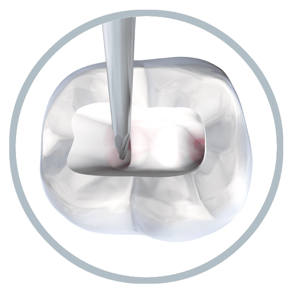
EndoGuard

EndoTracer

EndoExplorer
EndoGuard.
Perfecta visibilidad y seguridad.
La fresa EndoGuard pone desde el primer momento los medios para que el tratamiento endodóntico alcance el éxito deseado. Cualquier tratamiento endodóntico exitoso se basa en la creación de una cavidad de acceso perfecta. Por esta razón, comparado con la preparación del conducto radicular subsiguiente, esta fase de trabajo necesita muchas veces mayor tiempo de trabajo.
Utilizada inmediatamente después de crear el acceso a la cámara pulpar, la EndoGuard de Komet le ayuda a realizar este paso con gran eficacia y máxima seguridad
El curso está fijado para el éxito endodóntico. Tus ventajas
Cavidad de acceso perfecta
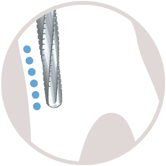
Después de crear el acceso la cámara pulpar, y remover la dentina excesiva con la EndoGuard, la visibilidad de la cámara pulpar mejora, y se facilita la detección de las entradas a los conductos radiculares. El acceso recto al sistema radicular minimiza el riesgo de transportaciones del conducto y fractura de la lima.
Protección del fondo de la cámara pulpar
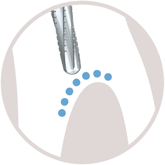
Dotada con una punta lisa inactiva, la EndoGuard es capaz de conservar el fondo de la cámara pulpar, y de evitar la remoción excesiva de sustancia sana.
No hay socavaduras
Gracias a su forma cónica, con la EndoGuard se evita la preparación involuntaria de socavaduras, asegurando así que no permanezcan residuos de tejido infectado dentro de la cavidad pulpar.
Dentadura efficiente con cortes transversales a lo largo de sus filos
La dentadura con cortes transversales a lo largo de sus filos de la EndoGuard permite trabajar de forma eficiente con el mayor control de trabajo en todo momento.
Un tratamiento exitoso y seguro del conducto radicular se basa en la creación de un acceso recto al sistema radicular.
Mientras la dentina protuberante en la parte distal del diente reduce la visibilidad del sistema radicular, gracias al uso de la EndoGuard la visibilidad de los conductos radiculares es excelente en la parte mesial.
EndoTracer.
Detecta todos los conductos con seguridad.
EndoTracer es un instrumento que sirve para la preparación del istmo. Por regla general los conductos radiculares de dientes con múltiples raíces no se detectan con facilidad, ni se puede penetrar en ellos inmediatamente. En muchos casos, hay que preparar un istmo a lo largo del trayecto o bien una parte del mismo , para exponer un conducto obliterado.
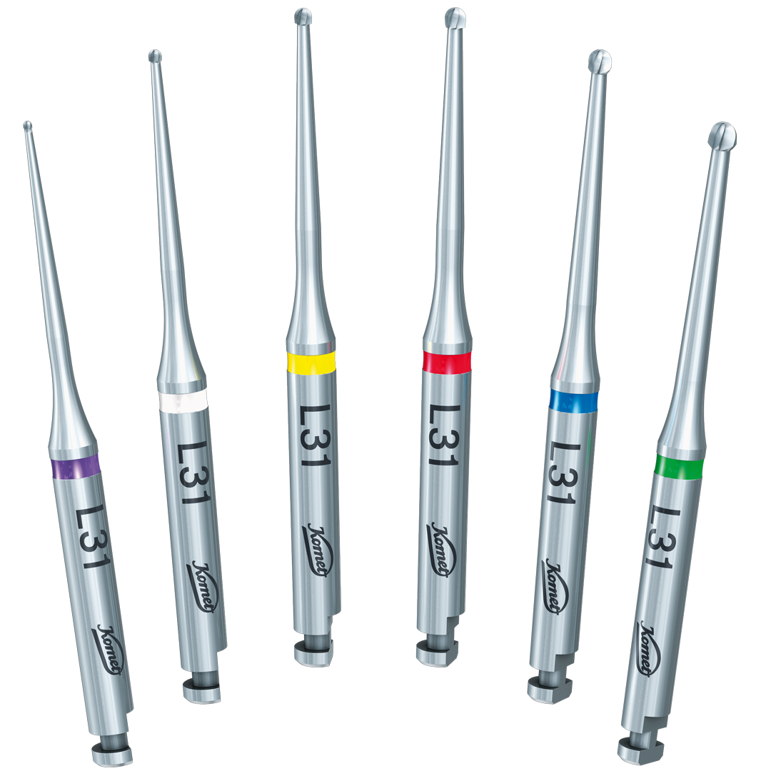
EndoTracer posee un largo y estrecho cuello, que asegura una buena visibilidad de la cavidad de acceso. Gracias a ello se pueden ver claramente las zonas más profundas de la cavidad, facilitando así la exposición del fondo de la cámara pulpar, la apertura conservadora de las entradas radiculares y la exposición de los conductos obliterados
EndoTracer. Tus ventajas.
Trabajo mínimamente invasivo
Las esbeltas fresas redondeadas, en especial los tamaños 004 y 006, son ideales gracias a su diseño para un trabajo fino de istmos y para las entradas radiculares, cumpliendo así con las exigencias de la endodoncia mínimamente ivasiva.
Ideal para su utilización con microscopio
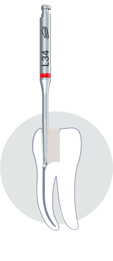
Gracias al largo y delgado cuello esta fresa permite una excelente visibilidad de la cavidad de acceso por delante del instrumento. Existe una versión del EndoTracer con una longitud total de 34 mm, ésta se ha adaptado de manera que la construcción de esta versión es ahora 3 mm más larga y por ello más adecuada para trabajos bajo el microscopio. En especial en el caso de largas coronas clínicas es cuando EndoTracer L34 demuestra sus ventajas
El tamaño correcto para cada diente
El EndoTracer está disponible en dos longitudes de 31 mm y 34 mm. Cada longitud viene en 6 tamaños: 004, 006, 008, 010, 012 y 014. De modo que para cada situación clínica existe el instrumento adecuado.
Dentadura afilada
EndoTracer posee una dentadura afilada, gracias a ella se puede eliminar la dentina y exponer la cavidad de acceso casi sin presión y de manera poco invasiva.
EndoExplorer.
Encuentra el camino con un dieseño innovador .
En los últimos años la odontología y en particular la endodoncia han experimentado un cambio paradigmático hacia tratamientos mínimamente invasivos. La creación conservadora de la cavidad de acceso y la creación de una apertura de trepanación pequeña permiten preservar al máximo la sustancia sana del diente. Con la minimización del riesgo de fracturas del diente y de la raíz, se ve considerablemente mejorado el pronóstico de éxito del tratamiento endodóntico a largo plazo.
Los instrumentos del set EndoExplorer no sólo están a la altura del momento, sino que también marcan nuevos estándares con gran rapidez. Específicamente diseñado para utilizar con un microscopio, el diseño innovador de estos instrumentos facilita la creación mínimamente invasiva de la cavidad del acceso endodóntico con un máximo control de trabajo.
No solo trabajamos con las últimas innovaciones, sino que las superamos. Tus ventajas
Todo a la vista
El diseño de los instrumentos se adapta perfectamente a las exigencias de los dentistas que trabajan con un microscopio. El delicado diseño de la parte activa del instrumento y el cuello largo y estrecho de éste permiten en cualquier momento una buena visibilidad del campo operatorio, con una ampliación de hasta 20x bajo el microscopio.
Menos presión, más eficacia
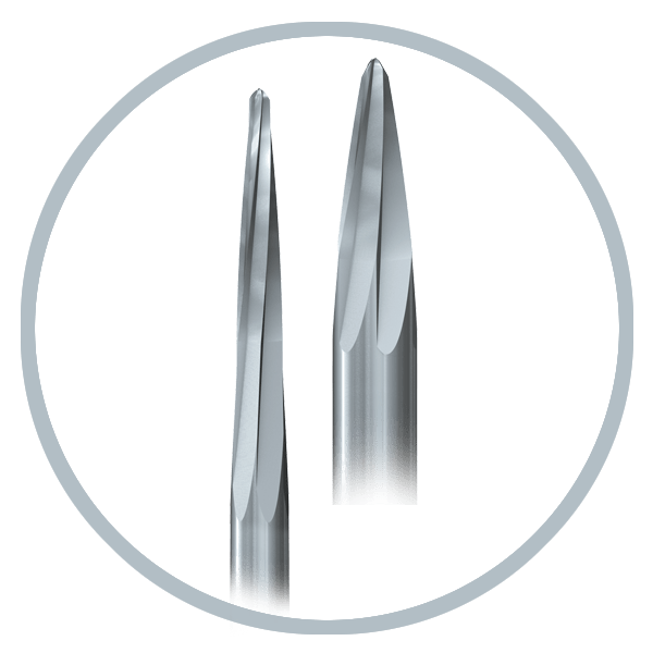
Los instrumentos EndoExplorer están dotados de una estructura dentada muy afilada. Esto permite retirar la sustancia dura del diente con precisión y casi sin presión, procurando un trabajo controlado y facilitando la creación eficiente de la cavidad del acceso endodóntico.
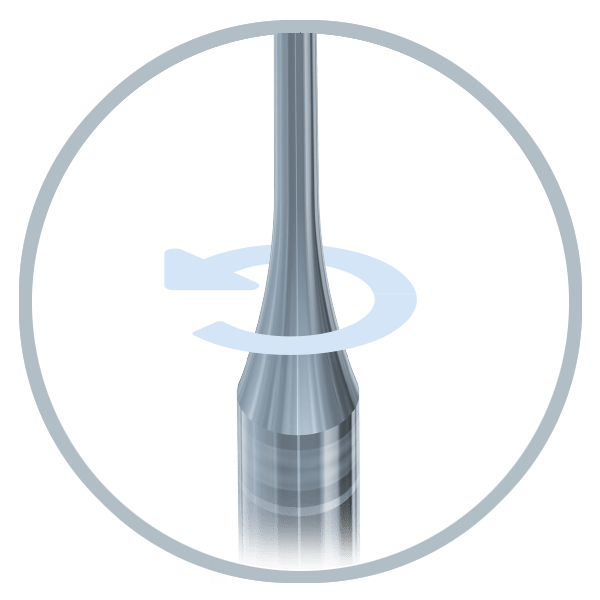
Perfecta marcha concéntrica
Los instrumentos EndoExplorer están fabricadosbcon carburo de tungsteno hasta el mango, esto garantiza una elevada concentricidad incluso después de haberse utilizado en numerosas ocasiones.
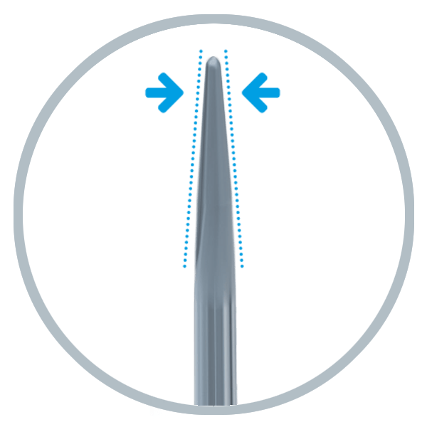
En nuevo enfoque
Gracias a la forma cónica de la parte activa del instrumento EndoExplorer permite una dirección controlada. De modo que la sustancia dura del diente puede retirarse con gran precisión y al mismo tiempo se conserva la valiosa dentina cervical. Este enfoque mínimamente invasivo mejora el pronóstico a largo plazo de los dientes tratados endodónticamente.
La llamada «Endodoncia mínimamente invasiva» parece ser la palabra de moda, pero a menudo no se tiene en cuenta que esto no se trata de una nueva demanda revolucionaria, es más el deseo de conservar un máximo la sustancia del diente algo que siempre se ha considerado como una de las metas principales de una odontología comprometida, sin embargo hasta ahora se carecía de posibilidades de poner este principio en la práctica de manera fiable. Gracias al nuevo set de instrumentos EndoExplorer, combinados con las fresas redondas H1SML y los medios de aumento óptico apropiados, el dentista dispone ahora del instrumental adecuado para poder crear el acceso endodóntico requerido con una máxima conservación de las sustancia, según la divisa «tan pequeño como sea posible , tan grande como sea necesario» y esto sin que el uso subsiguiente de los instrumentos endodónticos necesarios sea limitado o comprometido . Los dos tipos de instrumentos han logrado convertirse en una parte integral de nuestro proceso de trabajo endodóntico y constituyen una ayuda valiosa en nuestro trabajo cotídiano.
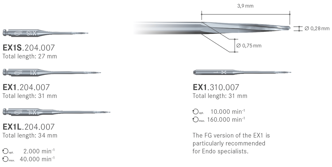
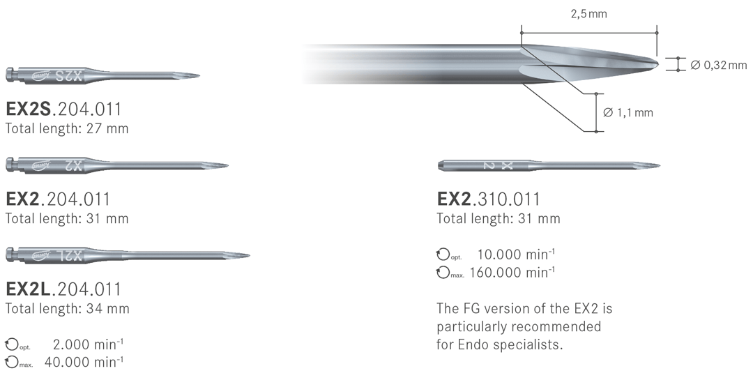
EndoExplorer componentes del sistema
Indicaciones del instrumento EX1:
- Exposición del fondo de la cámara pulpar
- Creación conservativa del acceso a las entradas del conducto de la raíz
- Exposición de los conductos radiculares obliterados
- Exposición de los fragmentos de postes o de instrumentos
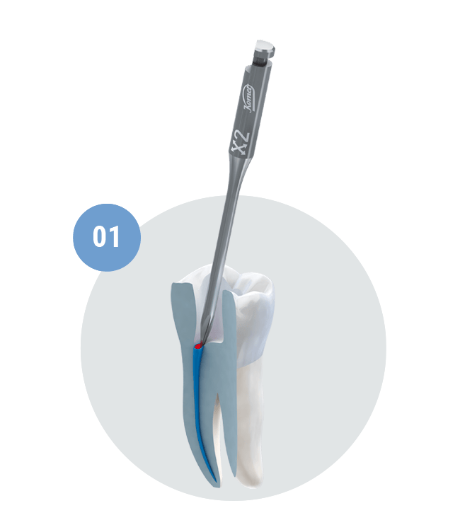
Paso a paso.
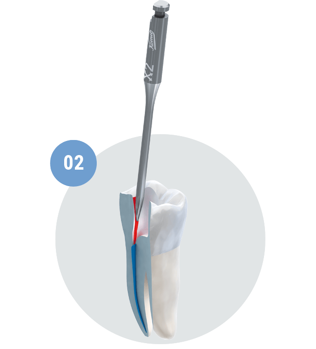
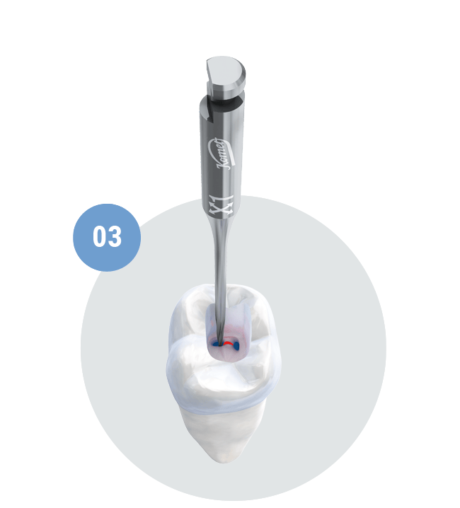
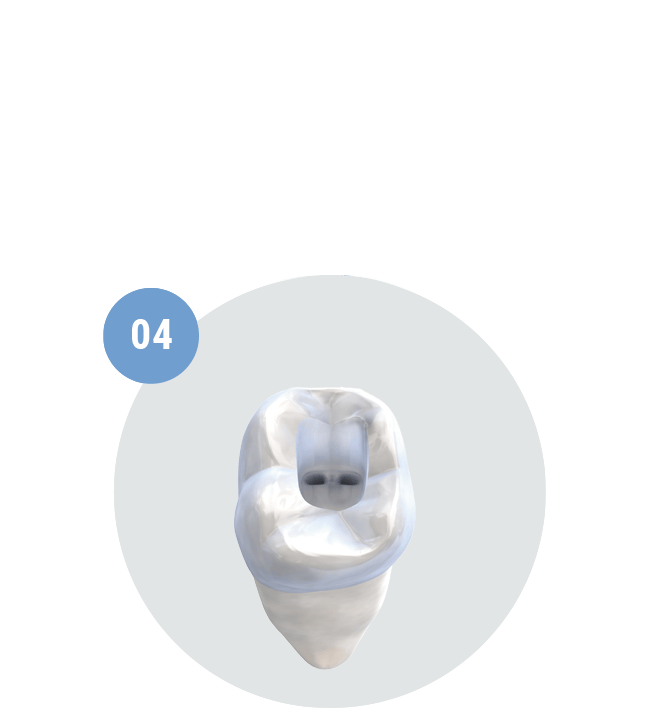
Formación inicial de la cavidad de acceso y exposición de las entradas a los conductos con el instrumento EX2.
Ensanchado de la cavidad de acceso con el instrumento EX2 moviéndolo en dirección al borde de la cavidad.

