Root canal treatment with Komet’s FQ file system
Root canal treatments are often the last resort for patients to preserve their natural teeth. As the procedure is part of the typical repertoire of general dentists, most patients are treated in general practices. Only in more advanced cases, patients are referred to specialized endodontists.
Komet’s FQ File System offers maximum flexibility that helps both general dentists and specialists like Dr. Rafaël Michiels to achieve perfect results. The expert from Hasselt, Belgium, kindly let us accompany him during some recent procedures, and we gained valuable insights and tips for the perfect endodontic treatment. While there is a basic protocol Dr. Michiels follows during every treatment, each patient has his or her own unique characteristics that affect the procedure – like in the two cases we discussed with Dr. Michiels. Read on to find out more about common challenges in endodontic treatments and how to conquer them.
Patient A: Pulpal necrosis and limited mouth opening

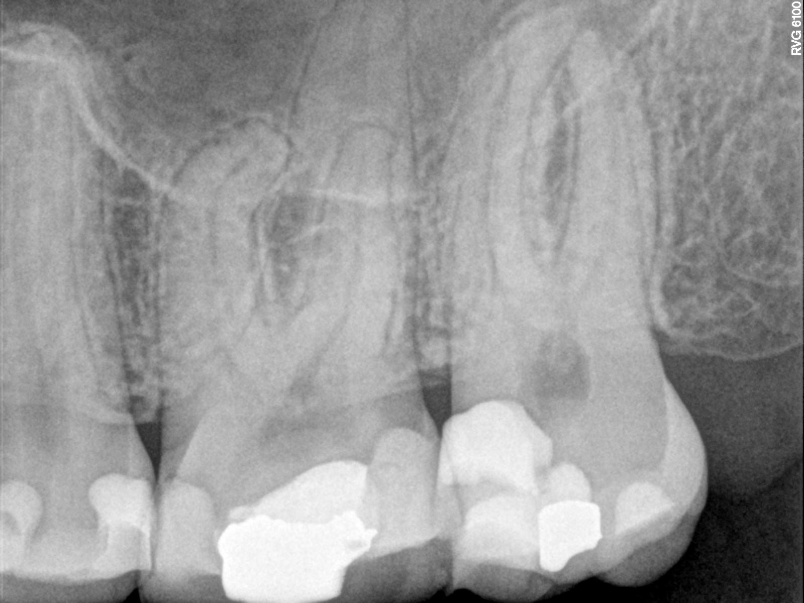
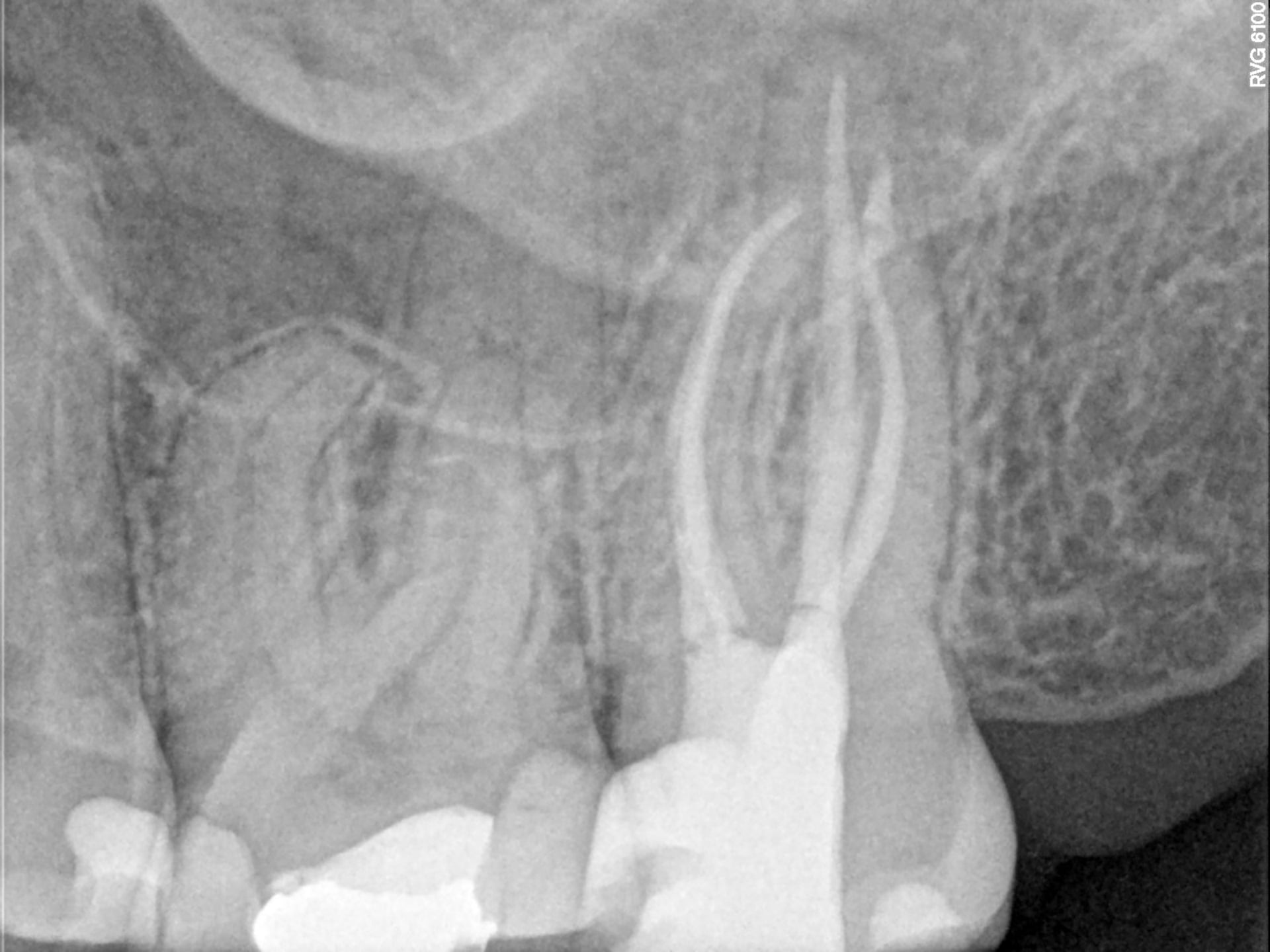
Radiograph of the teeth before the endodontic treatment
The first patient was initially referred to Dr. Michiels in June 2023. A deep restoration on the mesial side had been placed in a previous session and the attending dentist established that root canal treatment was required. In the first consultation, however, the patient had no complications and the tooth tested vital.
Only months later, the patient called with increasing pressure pain which led to the diagnosis of pulpal necrosis after deep restoration with acute symptomatic apical periodontitis. Hence, the root canal treatment was no longer an option.
As the infected tooth was the upper left second molar, the visualization of the tooth was already difficult. It was further compromised by the patient suffering from limited mouth opening, making the subsequent treatment even more challenging.
Patient B: Pulpal necrosis and a zirconia crown
As the patient had a zirconia crown on the affected tooth, he was referred to Dr. Michiels by his general dentist: Crowns are more difficult to open and also compromise the visualization of pulpal structures, the access cavity and in some cases the canals.
The radiographs which had been taken prior to the treatment revealed a further characteristic: The patient had calcified mesial canals, which makes them more difficult to detect once the crown has been opened.

Radiograph of the teeth before the endodontic treatment
Step-by-step: A deep dive into the endodontic treatment
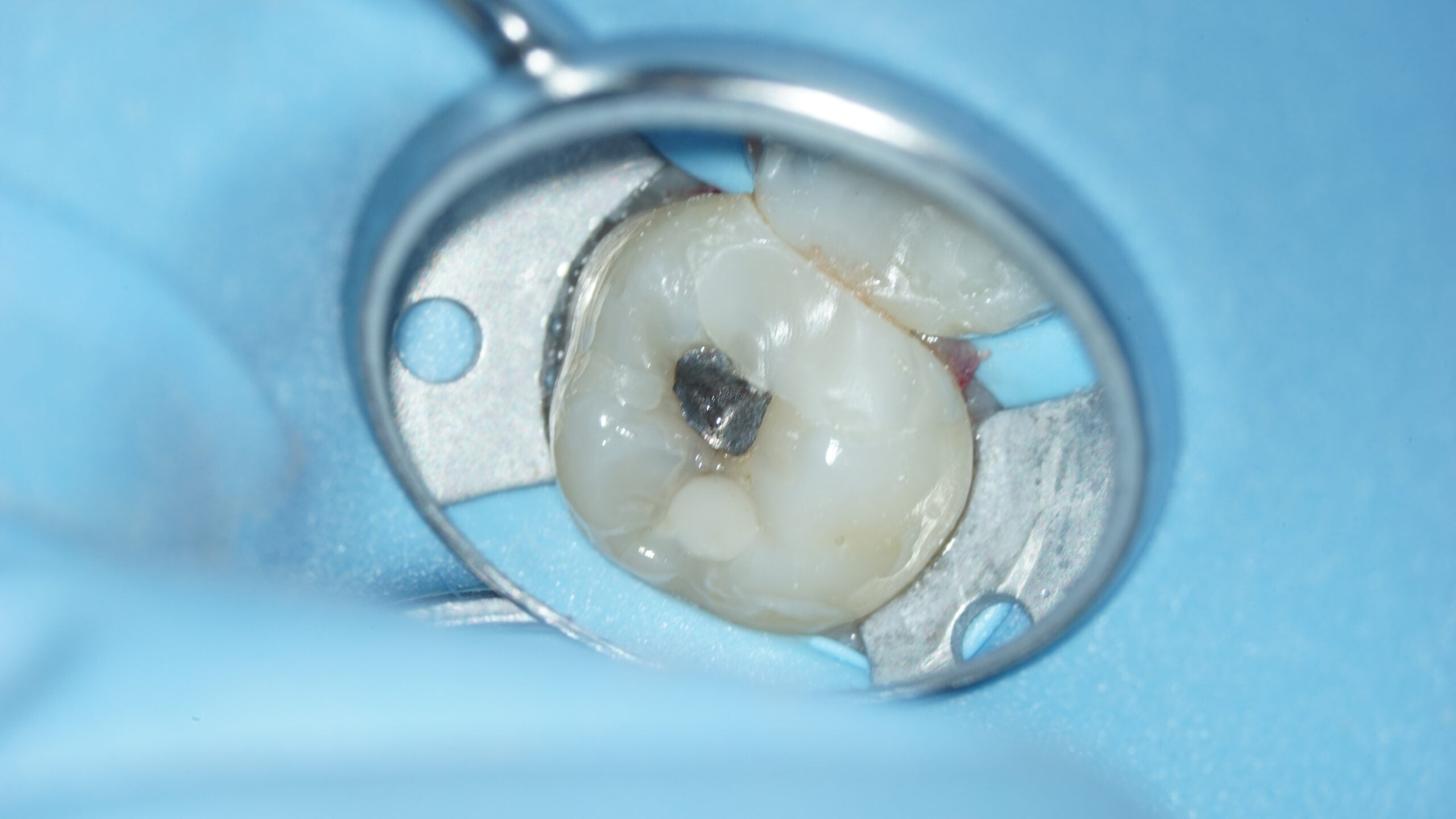
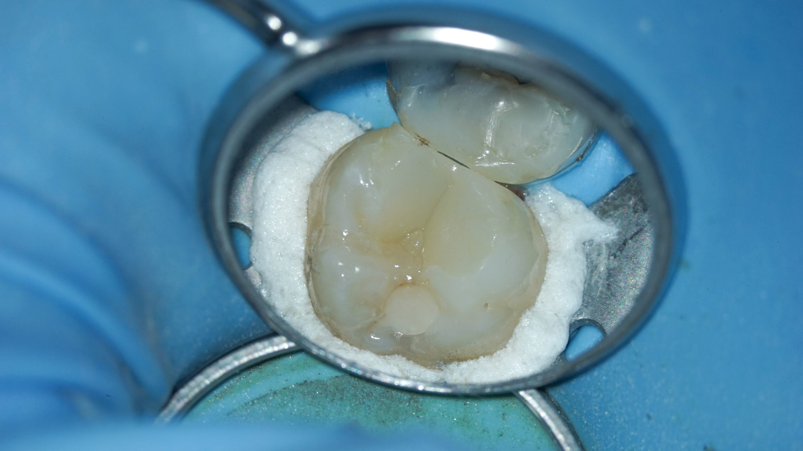
An endodontic treatment starts with the proper preparation of the treatment area. In order to prevent infection, Dr. Michiels chose to isolate the area by placing a rubber dam on several teeth. This method of isolating is particularly useful for patients with limited mouth opening, as it also increases the space to maneuver throughout the treatment.
Once the rubber dam was in place, Dr. Michiels created an opening cavity with Komet’s cylindrical bur 837KR. In addition, a combination of the Komet 851 Batt Bur and long neck bur was chosen to finish the access cavity.
The combination of these burs is ideal for any regular access cavity, as it allows easy opening and minimizes the risk of perforation. Of course, if your patient has a crown you have to adjust your choice of primary bur accordingly – otherwise, you will simply destroy your regular bur. For my patient’s zirconia crown, I used the Komet 4ZR 012.
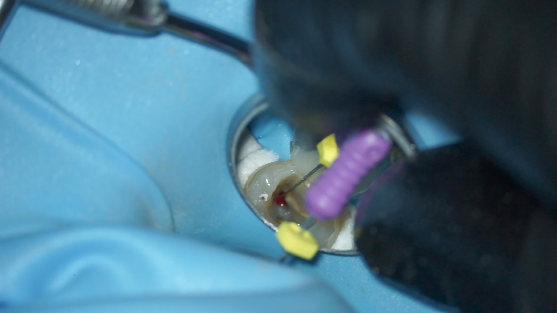
The FQ Opener was then used to locate and open the root canals, followed by hand files for working length determination. In the next step, Komet’s FQ Glider was used to create glide paths. As the high risk of blockage contraindicates the immediate use of regular instruments, creating glide paths is of the utmost importance.
However, if a patient has calcified mesial canals, you cannot go in with regular instruments straight away as this would increase the risk of fracture. Instead, Dr. Michiels advises to use small hand files, provided plenty of irrigation, and to gently create a glide path by hand before you switch to regular instruments.
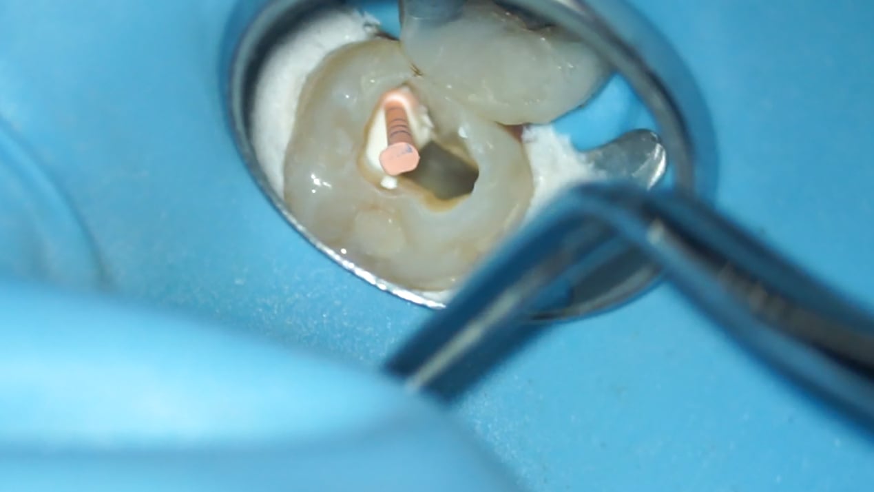
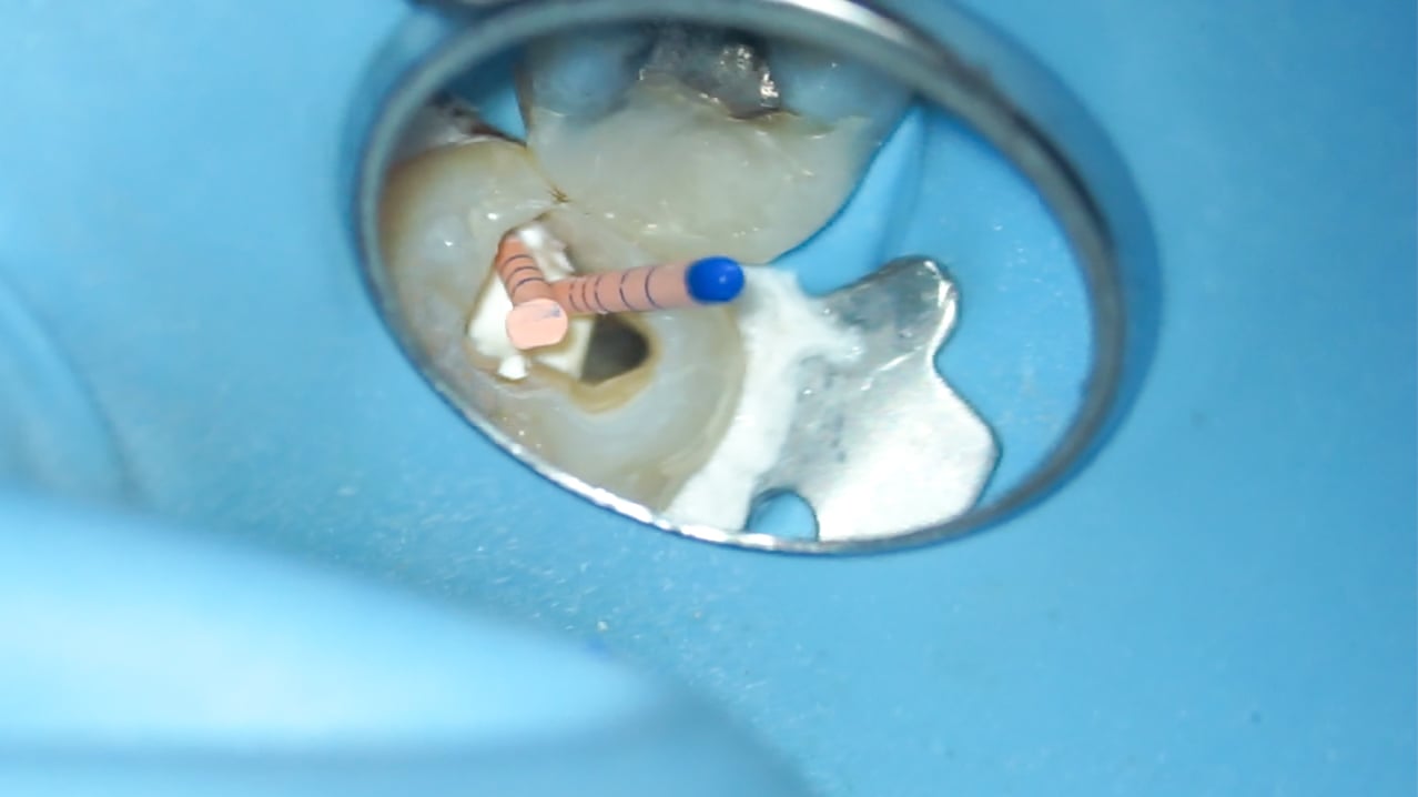
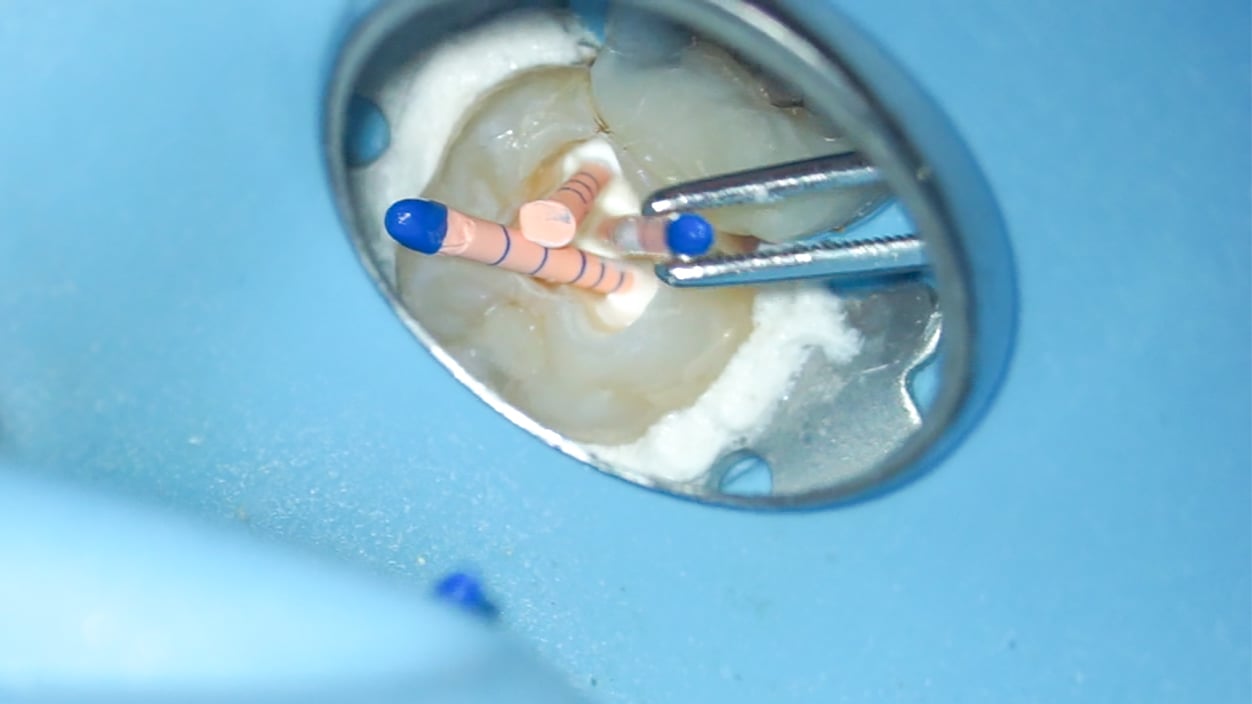
Once the glide paths were created, in this case with Komet’s FQ Glider, the FQ 025 file with taper .04 was used until it reached the working length, followed by the FQ file 035 with taper .04 to conclude the shaping of the root canals. The choice of taper used for this phase of the treatment was based on the subsequent obturation method. With a single cone obturation, the .04 taper is the more efficient solution as it helps to maintain as much of the tooth structure as possible. When using a warm obturation technique, however, the .06 taper is the better option.
Throughout the entire treatment, rinsing plays a key role. During the shaping of the root canals, sodium hypochlorite (5.25 %) was used for irrigation. At the end, the area was rinsed with citric acid (40 %) before the obturation was done according to the single-cone technique with Komet BioSeal, thus finishing the treatment in an efficient way.
It might not seem that crucial, but debris removal is very important for a successful treatment – especially if the patient has calcified canals. Residues of debris left in the canals lead to a blockage which makes irrigation significantly more difficult. Thanks to the variable taper of Komet’s FQ files, there is a noticeable difference compared to other instruments, and all debris is removed.
Modern endodontics: The impact of advanced file systems
While a root canal treatment is often considered to be a long and demanding procedure, advanced instruments and file systems enable general dentists to perform endodontic treatments at their own practice in a particularly efficient and comfortable way. This evolution of endodontic technologies allows general dentists to offer a wider range of services and comprehensive care that is tailored to their patients’ medical background and needs.
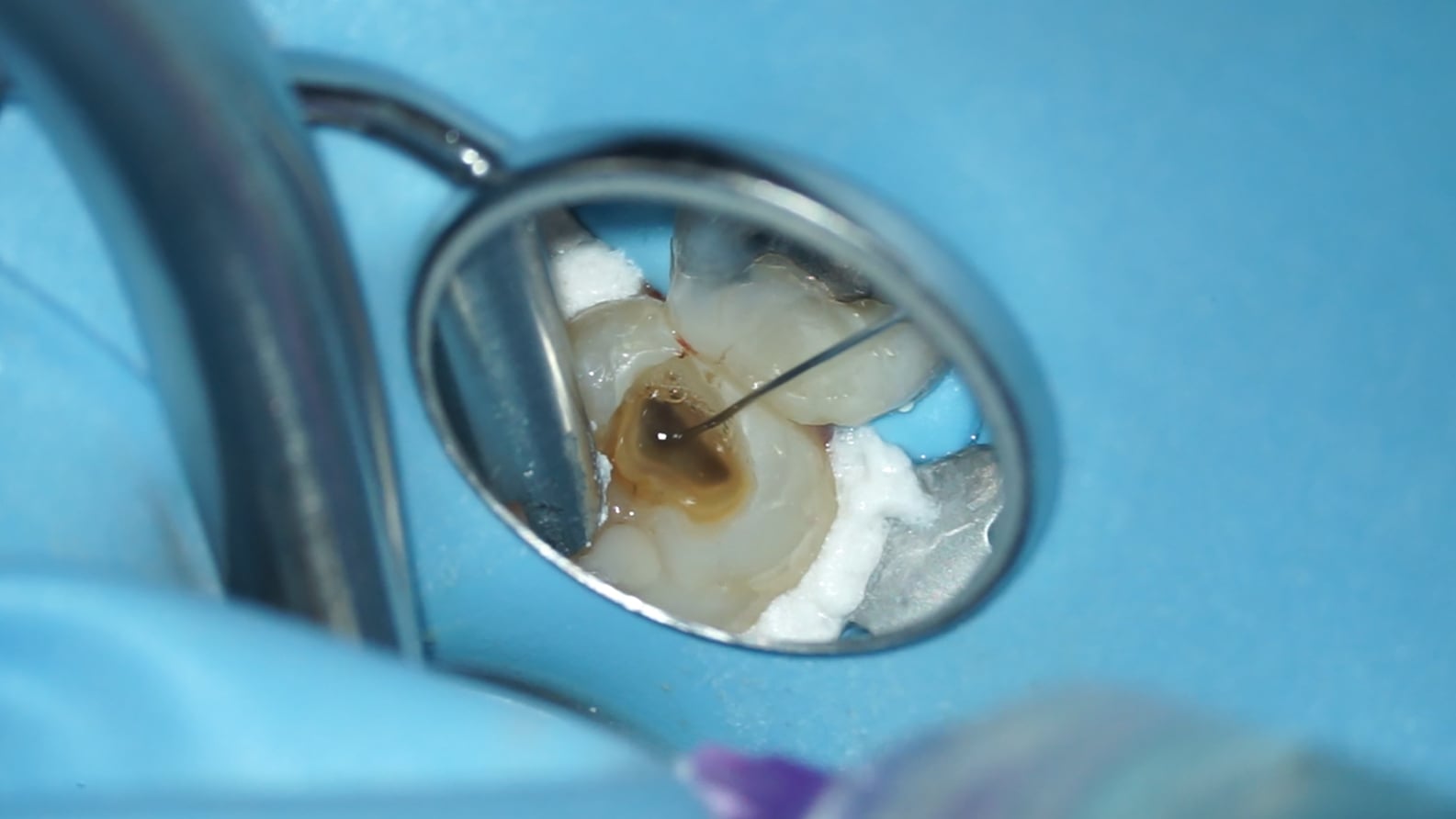
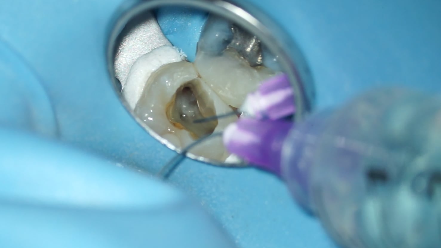
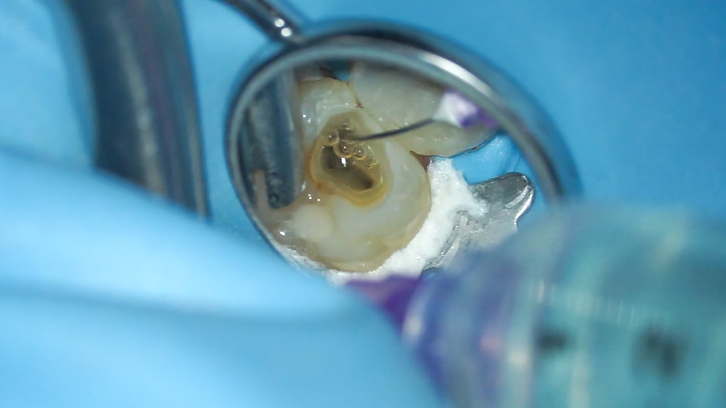
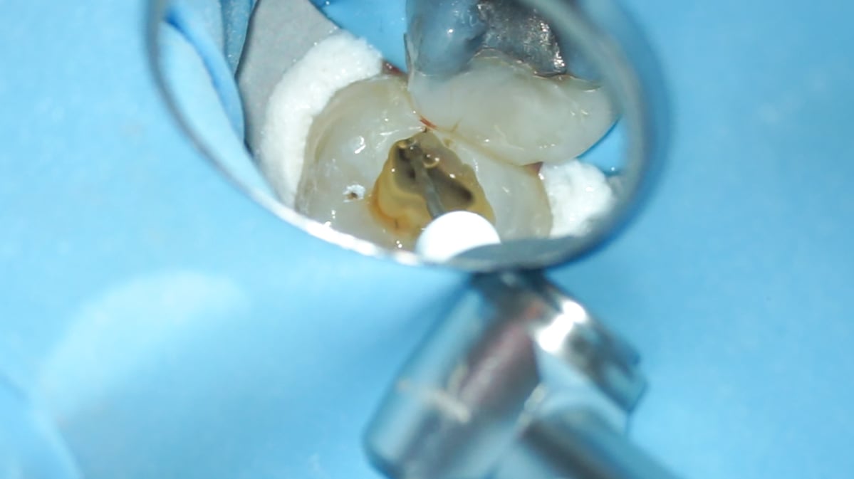
Inside the treatment room: A real-time look on the endodontic procedure
Dr. Rafaël Michiels performing a root canal treatment on the upper left second molar of his patient. (Patient A)
From dentist to dentist: 5 tips for your upcoming treatment
1. Isolation of multiple teeth.
Rubber dam isolation is often used on single teeth. During the actual treatment, however, it is helpful to isolate a quadrant or three or four teeth. In addition, attaching the clamp to a more distant tooth helps to improve visualization and gain more space to maneuver throughout the treatment.
2. Creating a straight access cavity.
It is very important to have straight access to the canals. Nowadays, the trend is to create access cavities that are as small as possible. But in cases where a patient suffers from limited mouth opening, this will further restrict the access to the canals. Thus, a standard access cavity will help you to achieve a better result.
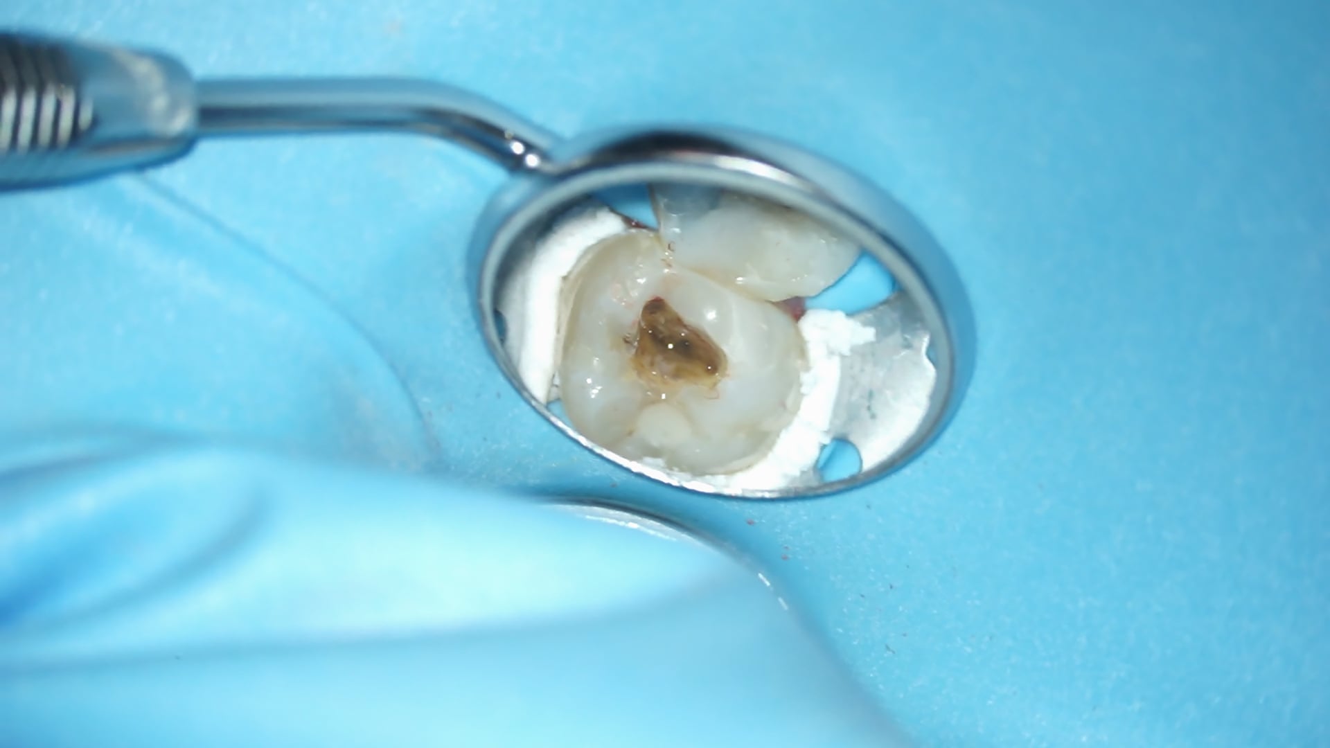
3. Detecting calcified canals.
Locating your patient’s root canals is an essential precondition for any endodontic treatment. However, some patients may have calcified canals, which makes them far more difficult to detect. The key is to take a closer look at the shade of the dentin. Calcified canals tend to be of a darker shade than the rest of the dentin. They can be opened by gently using a hand file or treated with citric acid.
4. Choosing the right instruments.
From special burs adapted to your patient’s type of crown to a flexible file system like FQ to support you in thorough debris removal: Choosing the right instruments for your treatment helps to reduce the time spent on the preparation, increase efficiency, and make the treatment more comfortable for your patients.
5. Investing in useful equipment.
Whether your patient suffers from limited mouth opening, has a crown and even if there are no special circumstances that need to be taken into account, illumination and visualization are always of the utmost importance when it comes to performing an endodontic treatment with optimum results. Thus, investing in a microscope, loupes with lighting or similar visual aids is essential, and often the equipment can be used for further cases as well.
About the dentist
Dr. Rafaël Michiels

Dr. Rafaël Michiels studied dentistry at the University of Ghent. After his graduation in 2006, he chose to specialize in endodontics and completed his postgraduate training in 2009. In 2013, Dr. Michiels started his own referral-based endodontic practice in Hasselt, Belgium, where he performs root canal treatments on numerous patients. He is a certified member of the European Society of Endodontology (ESE), Belgian Association for Endodontology and Traumatology (BAET) as well as the Dutch Association for Endodontology (NVvE) and has lectured at both national and international conferences.
